Leta i den här bloggen

måndag 4 juni 2018

(2) Hepoxiliinit Hx ja trioxiliinit TrX

(Wikipedialähteestä. Suomennosta Hepoksiliineistä) 

Historiasta.

Hepoxiliinit HsA3 ja HxB3 tunnistettiin jo vuonna 1984. Niiden havaittiin stimuloivan insuliinineritystä haimasaarekkeissa koe-eläimellä. Tämän havaitsi Pace-Asciak CR et Martin JM. Pace-Asciak myös tunnsiti, nimesi ja osoitti niiden insuliinisekretagogisen aktiivisuuden.

  • History HxA3 and HxB3 were first identified, named, shown to have biological activity in stimulating insulin secretion in cultured rat pancreatic islets of Langerhans in Canada in 1984 by CR Pace-Asciak and JM Martin.[1] Shortly thereafter, Pace-Asciak identified, named, and showed to have insulin secretagogue activity HxA4 and HxB4.[2]

Nomenclature

Hepoxiliinit

ARAKIDONIHAPPO (AA) JOHDANNAISET
Hepoxiliinit Hx (HXA3 ja HxB3 ovat muista eikosanoideista eroavia sikäli, että ne ovat muodosotuneet 20 hiilen rasvahapoista oksidoitumalla siten, että niissä on sekä epoksidi että hydroksiditähteitä. Rakenteellisesti ne eroavat erityisesti kahdesta muusta arakidonin eikosanoidiryhmästä LT ja LX (leukotrieenit ja lipoksiinit) siinä, että niistä puuttuu konjugoituneet kaksoissidokset. 
 Arakidonihapon ei-klassisista eikosanoideista tulee hepoxiliinejä 14,15-HxA3 ja 14,15-HxB3 (ultimate double bond, last double bond, viimeiseen kaksoissidokseen muodostunut epoksiryhmä). Niitä muodostuu toista metabolista tietä kuin mainitut klassisset eikosanoidit ja niiden hepoksiliinit.

  • The 14,15-HxA3 and 14,15-HxB3 non-classical eicosanoids are distinguished from the aforementioned hepoxilins in that they are formed by a different metabolic pathway and differ in the positioning of their epoxide and hydroxyl residues.
EIKOSAPENTAEENIHAPPO (EPA) JOHDANNAISET
Hepoxiliinit HxA4 ja HxB4 eroavat vastaavista arakidonihapon hepoksiliineistä siinä, että niissä on neljä kaksoissidosta (eikä kolme) jäljellä.
  • HxA4 and HxB4 are distinguished from HxA3 and HxB3 by possessing four rather than three double bonds.

Hepoxiliinin kaltaiset

DOKOSAHEXAEENIHAPON (DHA, C22:6 n3) hepoxiliinijohdannaiset 

LINOLIHAPON(LA, C18:2 n6) hepoxiliinin kaltainen johdannainen sfingosiini, esteröitynyt omega-hydroksyloitunut-sfingosiini (EOS)
  • Two other classes of epoxyalcohol fatty acids, those derived from the 22-carbon polyunsaturated fatty acid, docosahexaenoic acid, and the 18-carbon fatty acid, linoleic acid, are distinguished from the aforementioned hepoxilins by their carbon chain length; they are termed hepoxilin-like rather than hepoxilins.[3][4] A hepoxilin-like derivative of linoleic acid is formed on linoleic acid that is esterified to a sphingosine in a complex lipid termed esterified omega-hydroxylacyl-sphingosin (EOS).[4]

Note on nomenclature ambiguities

Kahdesta tärkeästä näkökohdasta hepoxiliinien ja niiden kaltaisten yhdisteiden rakenteelliset identiteetit useimmissa tutkimuksissa ovat epäselviä. Ensinnäkin niiden OH-ryhmien R/S-kiraalisuus alkututkimuksissa ja useassa myöhemmässä tutkimuksessa on määrittämätön ja sen takia ilmaistaan esm. HxB3n suhteen 10R/S-OH eli 10R/S hydroxy tai vain 10-OH eli 10-hydroxy.
 Toiseksi epoksidin R,S tai S,R kiraalisuus on määrittämättä ja ilmaistaan esim. HxB3:n suhteen 11,12-epoksidina. Muutamissa myöhemmissä tutkimuksissa on määritelty näiden tähteiden kiraalisuus, mutta ei ole varmaa, jos löytö on verrannollinen entisiin.
  • The full structural identities of the hepoxilins and hepoxilin-like compounds in most studies are unclear in two important respects. First, the R versus S chirality of their hydroxy residue in the initial and most studies thereafter is undefined and therefore given with, for example, HxB3 as 10R/S-hydroxy or just 10-hydroxy. Second, the R,S versus S,R chirality of the epoxide residue in these earlier studies likewise goes undefined and given with, for example, HxB3 as 11,12-epoxide. While some later studies have defined the chirality of these residues for the products they isolated,[5] it is often not clear that the earlier studies dealt with products that had exactly the same or a different chirality at these residues.

Biokemiaa, Biochemistry

Tuotanto

ARAKIDONIHAPPOPERÄISET HEPOXILIINIT (Hx3)
Ihmisen arakidonihappoperäiset hepoxiliinit (HxA3, HxB3) muodostuvat kahden vaiheen reaktioissa.
(1) Molekulaarinen happi O2 vaikuttaa yhteen kaksoissidokseen (Z12) ja toisessa kohtaa siirtyy kaksoissidos ja muodostuu hydroperoksidijohdos 12S-HpETE. 
2) 12S-HpETE konvertoituu hepoxiliiniksi HxA3 (Kuva Wikipediassa)
 IUPAC nimi on aika hankala, rakenne ilmaistaan pätkä kerrallaaa sen nimityksen tarkkuuden esiinsaamiseksi.
Toinen reaktio, jossa HpETE muoto muuttuu hepoxiliieniksi HxA3 tai HxB3 , saattaa riippuva entsyymistä ALOX12.
Poistogeenisiin tutkimuksiin perustuen ALOXE3, epidermaalinen lipoxygenaasi, vastaisi ainakin hiiren ihossa ja spinaalisessa kudoksessa 12(S)-HPETE muodon konversioista HxB3- muotoon.

Kudoksen 12(S)-HPETE muuttunee ei - entsymaattisestikin muotoihin HxA3 ja HxB3. Ei-entsymaattinen hepoxiliinimuodostus saattaa olla artefaktaa.
Lopuksi solun peroksidaasit vähentävät helposti ja nopeasti 12(S)-HPETE-muotoa sen hydroksi-analogiksi 12(S)-HETE, 12-hydroksieikosatetraeenihappo. Tämä reaktio kilpailee hepoxiliinien muodostumisen kanssa soluissa, joissa peroksidaasiaktiivisuus on hyvin suuri ja vastannee hepoxiliinituotannon blokeeraamisesta.
Entsyymi LOX15 vastaa arakidonihapon metaboloimisesta 14,15-hepoxiliiniA3:ksi . Tässä tiessä , jossa ultimate (last double bond) on muuntunut, on seuraavia tapahtumia. Ensin muodostuu 15-asemaan hydroperoksidi ja tulee 15(S)-HpETE ja siitä   kaksi  hepoksiliini-isomeeriä 14,15-HxA ja 14,15-HxB.

ALOX15 pystyy tekemään kummatkin reaktiovaiheet, vaikka jatkotutkimukset voivatkin osoittaa, että ALOXE3 ja ei-entsymaattiset uudelleenjärjestäytymiset  sekä  15(S)-HPETE:n redusoituminen vastaavan 15(S) HETE- muotoon saattavat olla osallisia  tuotteissa 14,15-HxA3 ja 14,15-HxB3 ( ultimate asema).

      • Production
  • Human HxA3 and HxB3 are formed in a two-step reaction. 
     (1) First, molecular oxygen (O2) is added to carbon 12 of arachidonic acid (i.e. 5Z,8Z,11Z,14Z-eicosatetraenoic acid) and concurrently the 8Z double bond in this arachidonate moves to the 9E position to form the intermediate product, 12S-hydroperoxy-5Z,8Z,10E,14Z-eicosatetraenoic acid (i.e. 12S-hydroperoxyeicosatetraenoic acid or 12S-HpETE).
    (2)  Second, 12S-HpETE is converted to the hepoxilin products, HxA3 (i.e. 8R/S-hydroxy-11,12-oxido-5Z,9E,14Z-eicosatrienoic acid) and HxB3 (i.e. 10R/S-hydroxy-11,12-oxido-5Z,8Z,14Z-eicosatrienoic acid).[3] 
    This two-step metabolic reaction is illustrated below:
     5Z,8Z,11Z,14Z-eicosatetraenoic acid + O2 
     → 12S-hydroperoxy-5Z,8Z,10E,14Z-eicosatetraenoic acid
     → 8R/S-hydroxy-11,12-oxido-5Z,9E,14Z-eicosatrienoic acid 
    +  10R/S-hydroxy-11,12-oxido-5Z,8Z,14Z-eicosatrienoic acid.

    The second step in this reaction, the conversion of 12(S)-HpETE to HxA3 and HxB3, may be catalyzed by ALOX12 as an intrinsic property of the enzyme.[6] 

     Based on gene knockout studies, however, the epidermal lipoxygenase, ALOXE3, or more correctly, its mouse ortholog Aloxe3, appears responsible for converting 12(S)-HpETE to HxB3 in mouse skin and spinal tissue.[4][7][8] 

     It is suggested that ALOXE3 contributes in part or whole to the production of HxB3 and perhaps other hepoxilins by tissues where it is expressed such as the skin.[4][9] 

     Furthermore, hydroperoxide-containing unsaturated fatty acids can rearrange non-enzymatically to form a variety of epoxyalcohol isomers.[10] 

     The 12(S)-HpETE formed in tissues, it is suggested, may similar rearrange non-enzymatically to form HxA3 and HXB3.[4] 

     Unlike the products made by ALOX12 and ALOXE3, which are stereospecific in forming only HxA3 and HxB3, however, this non-enzymatic production of hepoxilins may form a variety of hepoxilin isomers and occur as an artifact of tissue processing.[4] 

     Finally, cellular peroxidases readily and rapidly reduce 12(S)-HpETE to its hydroxyl analog, 12S-hydroxy-5Z,8Z,10E,14Z-eicosatetraenoic acid (12S-HETE; see 12-hydroxyeicosatetraenoic acid; this reaction competes with the hepoxilin-forming reaction and in cells expressing very high peroxidase activity may be responsible for blocking the formation of the hepoxilins.[3
  • ALOX15 is responsible for metabolizing arachidonic acid to 14,15-HxA3 and 14,15-HxB3 as indicated in the following two-step reaction which first forms 15(S)-hydroperoxy-5Z,8Z,11Z,13E-eicosatetraenoic acid (15S-HpETE) and then two specific isomers of
     11S/R-hydroxy-14S,15S-epoxy-5Z,8Z,12E-eicosatrienoic acid (i.e. 14,15-HxA3) and 
     13S/R)-hydroxy-14S,15S-epoxy-5Z,8Z,11Z-eicosatrienoic acid (i.e. 14,15-HxB3): 

    5Z,8Z,11Z,14Z-eicosatetraenoic acid + O2 
     → 15(S)-hydroperoxy-5Z,8Z,11Z,13E-eicosatetraenoic acid
     → 11R-hydroxy-14S,15 S-epoxy-5Z,8Z,12E-eicosatrienoic acid 
    and 13R-hydroxy-14S,15S-epoxy-5Z,8Z,11Z-eicosatrienoic acid
  • ALOX15 appears capable of conducting both steps in this reaction[11] although further studies may show that ALOXE3, non-enzymatic rearrangements, and the reduction of 15S-HpETE to 15(S)-hydroxy-5Z,8Z,11Z,13E-eicosatetraenoic acid (i.e. 15S-HETE; see 15-hydroxyicosatetraenoic acid) may be involved in the production of 14,15-HxA3 and 14,15-HxB3 as they are in that of HxA3 and HxB3.
DOKOSAHEXAEENIHAPPO (DHA, C22:6 n3), Hepoxiliinin kaltaiset metaboliittit

Hepoxiliinin kaltaisia metaboliitteja DHA:sta pääteltiin niiden trioksiliinimetaboliitteista (Trx) eli trihydroksimetaboliiteista käsin - näitä oli tuloksena, kun dokosahexaeenihappoa lisättiin rotasta saatuihin käpylisäke- tai hippokampi-isolaateihin. (DHA on aivoissa hyvin tavallinen rasvahappomuoto): 
7-OH-bis-alfa-dihomo-HxA5
10-OH-bis-alfa-dihomo-HxA5
  • Production of the hepoxilin-like metabolites of docosahexaenoic acid,
     7R/S-hydroxy-10,11-epoxy-4Z,7E,13Z,16Z,19Z-docosapentaenoic acid 
     (i.e. 7-hydroxy-bis-α-dihomo-HxA5) and
     10-hydroxy-13,14-epoxy-4Z,7EZ,11E,16Z,19Z-docosapentaenoic acid 
     (i.e. 10-hydroxy-bis-α-dihomo-HxA5)
     was formed (or inferred to be formed based on the formation of their trihydroxy metabolites (see trioxilins, below) as a result of adding docosahexaenoic acid to the pineal gland or hippocampus isolated from rats; the pathway(s) making these products has not been described.[3][12]

LINOLIHAPPO (LA, C18:2 n6), hepoxiliinien kaltaiset metaboliitit

Sekä ihmisellä että jyrsijöillä muodostuu ihossa/nahkassa hepoxiliinien kaltaisia metaboliitteja. Tämä hepoxiliini on esteröitynyttä sfinganiinia lipidikompleksissa (EOS, esteröitynyttä omega-OH-asyylisfingosiinia). Nämä asyylit eli rasvahappotähteet, joita sfinganiini ottaa esterisidoksilla vastaan voivat olla hyvin pitkiä rasvahappoja ( VLCFA, kuten aivojen sfingomyeliinissä).
Tässä metabolisessa tiessä ALOX12B metaboloi esteröityneen linolihapon 9R-hydroperoksijohdannaiseksi ja sitten ALOXE3 metaboloi tämän aineenvaihdunnallisen välituotteen 13-hydroxy-9,10-epoxituotteeksi. Tästä aineenvaihdunnallisesta tiestä ihoon saadaan asettumaan  hyvin pitkäketjuisia rasvahappoja ihon pinnan sarveistuneeseen lipidivaippaan.
  • A hepoxilin-like metabolite of linoleic acid forms in the skin of humans and rodents. This hepoxilin is esterified to sphinganine in a lipid complex termed EOS (i.e. esterified omega-hydroxyacyl-sphingosine, see Lipoxygenase#Biological function and classification#Human lipoxygenases) that also contains a very long chain fatty acid. In this pathway, ALOX12B metabolizes the esterified linoleic acid to its 9R-hydroperoxy derivative and then ALOXE3 metabolizes this intermediate to its 13R-hydroxy-9R,10R-epoxy product. The pathway functions to deliver very long chain fatty acids to the cornified lipid envelope of the skin surface.[9]

    Kommenttini:  Harper 1969: Tiedettiin jo 1958 linolihapon merkitys ihovaurioita parantavana ja essentiellinä rasvahappona. Samoin tiedettiin että linolihapon konversiossa arakidonihapoksi tarvittiin B6 vitamiinia , kuten  palmitiinihapon konversiossa sfingosiiniksi.
  • Harper jo 1969 mainitsee joitain aivojen tyypillisiä VLCFA rasvahappoja C24 lignoseriinihappo C24:0), nervonihappo 8C24:1), cerebronihappo (2-OH-C24:0), joita liittyy sfingosiiniin kerebrosideissa (sfingomyeliinin osissa). Laajentunut käsitys esentiellien rsvahappojen metaboliiteista on viime vuosikymmenien tietoa. Katabolinen tiekin on asia sinänsä.

Aineenvaihdunnan jatkoa

Hepoxiliini A3 on äärimmäisen epästabiili ja hepoxiliini B3 on kohtalaisen epästabiili ja ne muuttuvat nopeasti vastaaviksi trihydroksituotteikseen (TrX), kuten esim. kemiallisten isolaatioprosessien aikana jo lievästäkin happamuudesta  metodissa. Ne metaboloituvat nopeaati myös soluissa näiksi samoiksi trihydroksy-tuotteikseen, joita nimitetään trioksiliineiksi(Trx) tai trihydroksieikosatrieenihapoiksi (THETA).
Hepoksiliini A3 muuttuu trioksiliini A3:ksi (TrXA3).
Hepoksiliini B muuttuu trioksiliini B3:ksi (TrXB3).
Kolmas trioksiliinihappo arakidonihappolinjasta on trioksiliini C (TrXC), jota  on havaittu koe-eläimen aorttakudoksesta arakidonihappoinkubaatiossa.
 sEH-entsyymi, liukoinen epoksidihydrolaasi, vaikuttaa HxA3:n muuttumisen TrXA3:ksi ja HxB3:n muuttumisen TrX muotoon hiiren maksassa. Tätä entsyymiä on laajalti kudoksissa myös ihmisellä ja se voi olla pääasiallinen metaboloiva entsyymi näille ja muillekin hepoxiliinituotteille.

Hepoxiliinihydrolaasiaktiivisuutra voi olla muillakin entsyymeillä kuten mikrosomaalisella epoxidihydrolaasilla tai epoxidihydrolaasi 2:lla.

 Hepoxiliinituotannon trihydroksituotteet katsotaan inaktiiveiksi ja SEH-tie pidetään sen takia hepoxiliineja rajoittavana tienä, mutta kuitenkin on havaittu, että TrXA3, TRXB3 ja TRXC3 olivat koe-eläimessä hepoxiliinia HxA3 vahvempia supistetun arterian relaksoijia. Samoin TrXC3 relaksoi suhteellisen vahvasti supistunutta aorttaa. 

HxA3 pystyttiin muuttamaan glutationikonjugaatikseen HxA3-C, 11-glutationyyli-HxA3, soluttomassa systeemissä tai hippokampihomogenaatissa. Tämä HxA3-C osoittautui olevan vahva kalvon hyperpolarisaation stimuloija hippokampin CA1-neuroneissa. Tämä on LTC4 analogi ;( LTC4  on leukotrieeni LTA4, johon on konjugoitunut glutationi).

 On havaittu 14,15-HxA3 ja 14,15-HxB3 glutationikonjugaatteja Hodginin taudin Reed- Sternberg-solulinjassa.

 HxB3 ja TrX3 ovat olleet esteröityneinä sn2-asemaan ihmisen psoriasisleesion fosfolipidissä ja psoriaattiset ihonäytteet asyloivat HxB.. ja TrX.. näihin fosfolipideihin koeputkessa. ( Tässä ei ole selitystä, mitä nämä HxBw ja TrX2 tarkoittavat tai ovatko w ja 2  painovirhe).
      • Further metabolism
  • HxA3 is extremely unstable and HxB3 is moderately unstable, rapidly decomposing to their tri-hydroxy products, for example, during isolation procedures that use an even mildly acidic methods; they are also rapidly metabolized enzymatically in cells to these same tri-hydroxy products, termed trioxilins (TrX's) or trihydroxyeicoxatrienoic acids (THETA's); HxA3 is converted to 8,11,12-trihydroxy-5Z,9E,14Z-eicosatrienoic acid (trioxilin A3 or TrXA3) while TxB3 is converted to 10,11,12-trihydroxy-5Z,8Z,14Z-eicosatrienoic acid (trioxilin B3 or TrXB3).[3][13] A third trihydroxy acid, 8,9,12-trihydroxy-5Z,10E,14Z eicosatrienoic acid (trioxilin C3 or TrXC3), has been detected in rabbit and mouse aorta tissue incubated with arachidonic acid.[5][14] The metabolism of HxA3 to TrXA3 and HXB3 to TrX is accomplished by soluble epoxide hydrolase in mouse liver; since it is widely distributed in various tissues of various mammalian species, including humans, soluble epoxide hydrolase may be the principal enzyme responsible for metabolizing these and perhaps other hepoxilin compounds.[3][15] It seems possible, however, that other similarly acting epoxide hydrolases such as microsomal epoxide hydrolase or epoxide hydrolase 2 may prove to hepoxilin hydrolase activity. While the trihydroxy products of hepoxilin synthesis are generally considered to be inactive and the sEH pathway therefore considered as functioning to limiting the actions of the hepoxilins,[3][16] some studies found that TrXA3, TrXB3, and TrXC3 were more powerful than HxA3 in relaxing pre-contracted mouse arteries[5] and that TrXC3 was a relatively potent relaxer of rabbit pre-contracted aorta.[14]
  • HxA3 was converted through a Michael addition catalyzed by glutathione transferase to its glutathione conjugate, HxA3-C, i.e., 11-glutathionyl-HxA3, in a cell-free system or in homogenates of rat brain hippocampus tissue; HxA3-C proved to be a potent stimulator of membrane hyperpolarization in rat hippocampal CA1 neurons.[17] This formation of hepoxilin A3-C appears analogous to the formation of leukotriene C4 by the conjugation of glutathione to leukotriene A4. Glutathione conjugates of 14,15-HxA3 and 14,15-HxB3 have also been detected the human Hodgkin disease Reed–Sternberg cell line, L1236.[11]
  • HxB3 and TrX3 are found esterified into the sn-2 position of phospholipid in human psoriasis lesions and samples of human psoriatic skin acylate HxBw and TrX2 into these phospholipids in vitro.[3][18]

Fysiologiset vaikutukset

Miltei kaikki biologiset tutkimukset hepoxiliineistä on tehty eläimissä tai koeputkessa eläin- tai ihmiskudoksista. Kuitenkin näsitä tutkimuksista saa lajispesifisiä erovia tuloksia, mikä komplisoi asian relevanssin ihmiselle. Näiden tutkimusten hyöty ihmisfysiologiaan, patologiaan ja kliiniseen lääketieteeseen ja terapiatasoon vaatii paljon lisätutkimusta.

  • Physiological effects
    Virtually all of the biological studies on hepoxilins have been conducted in animals or in vitro on animal and human tissues, However, these studies give species-specific different results which complicate their relevancy to humans. The useful translation of these studies to human physiology, pathology, and clinical medicine and therapies requires much further study.

Tulehdus

HxA3 ja HxB3 omaavat proinflammatorisia vaikutuksia, esimerkiksi ne stimuloivat neutrofiilien kemotaxista ja lisäävät ihokapillaarien permeabiliteettia. Ihmisillä tehdyt tutkimukset ovat osoittaneet, että HxB3-pitoisuus on yli 16 kertaa korkeampi psoriaattisissa ihovaurioissa kuin normaalissa epidermiksessä – psoriasishilseessä sitä oli noin 10 mikromoolin pitoisuuksissa, millä on biologista vaikutusta. Varsinaista HxB3- muotoa ei näissä kudoksissa esiintynyt, vaan se pääteltiin metaboliitistaan TxB3, jota oli suhteellisen korkeat pitoisuudet psoriasishilseessä, mutta ei normaalissa epidermiksessä. Nämä tulokset viittaavat siihen, että HxA3 ja HxB3 hepoxiliinien proinflammatoriset vaikutukset voivat antaa osansa psoriasista seuraavaan tulehdusvasteeseen ja ehkä muihinkin tulehduksellisiin ihotiloihin.

HxA3 on osallistunut jyrsijäsuolistossa ja keuhkossa eri bakteerien aiheuttamaan neutrofiilipohjaiseen tulehdusvasteeseen. Tästä mahdollistuu tämän hepoxiliinin edistävä vaikutus muidenkin kudosten tulehdusvasteisiin, erityisesti limakalvopintaisissa kudoksissa ihon lisäksi.

 Lisäksi HxA3 ja HxB3:n synteettinen analogi PBT-3 indusoivat ihmisen neutrofiilejä tuottamaan neutrofiilin extrasellulaarista verkostoa, nimittäin DNA-pitoista solunulkoista säiematriisia, histonipitoista kromatiinia, joka kykenee tappamaan solun ulkopuolisia patogeenejä minimoimalla kudosta; näin nämä hepoxiliinit voivat ottaa osaa luonnolliseen immuniteettiin vastaamalla patogeenien suorasta tuhoamisesta.
  • Inflammation
  • HxA3 and HxB3 possess pro-inflammatory actions in, for example, stimulating human neutrophil chemotaxis and increasing the permeability of skin capillaries.[3][19] Studies in humans have found that the amount of HxB3 is >16-fold higher in psoriatic lesions than normal epidermis. It is present in psoriatic scales at ~10 micromolar, a concentration which is able to exert biologic effects; HxB3 was not detected in these tissues although its present was strongly indicated by the presence of its metabolite, TrXB3, at relatively high levels in psoriatic scales but not normal epidermal tissue.[13] These results suggest that the pro-inflammatory effects of HxA3 and HxB3 may contribute to the inflammatory response that accompanies psoriasis and perhaps other inflammatory skin conditions.[3][13][20][21] HxA3 has also been implicating in promoting the neutrophil-based inflammatory response to various bacteria in the intestines and lungs of rodents.;[22][23] this allows that this hepoxilin may also promote the inflammatory response of humans in other tissues, particularly those with a mucosa surface, besides the skin. In addition, HxA3 and a synthetic analog of HxB3, PBT-3, induce human neutrophils to produce neutrophil extracellular traps, i.e. DNA-rich extracellular fibril matrixes able to kill extracellular pathogens while minimizing tissue; hence these hepoxilins may contribute to innate immunity by being responsible of the direct killing of pathogens.[24]

Verenkierto

Sekä 12S-HETE ja 12R-HETE että HxA3 ja TrXA3 vaikuttavat hiiren tromboksaani A2:lla (TXA2) supistetuissa mesenteriaalivaltimoissa relaksaatiota, mutta tätä vaikutusta ei saatu esiin HxB3 eikä TrX3:lla.
 Mekanistisesti näitä metaboliitteja muodostuu suoniendoteelissä, niitä liikkuu alla olevaan sileään lihakseen ja ne purkavat reseptoriantagonisteina sileän lihaksen supistustilan, jonka TXA2 on saanut aikaan vaikuttamalla reseptoriinsa (tromboxaanireseptori alfaisomeeri).
Arakidonihapon 15-LOX johdannaiset epoksialkoholi- ja TrX-metaboliitit kuten
15-hydroxy-11,12-epoxyeikosatrieenihappo,
13-hydroksy-14,15-epoxy-eikosatrieenihappo ( 14,15 HxA4 isomeeri EPA- rasvahaposta?)
11,12,14- trihydroksieikosatrieenihappo arakidonihaposta
laajentavat kaniinin aorttaa EDHF-mekanismilla eli endoteeliperäisellä hyperpolarisoivalla tekijällä. Ne muodostuvat suonen endoteelissä, liikkuvat allaolevaan sileään lihakseen ja liipaisevat esiin hyperpolarisaation indusoiman relaksaation sitoutumalla kalsiumilla aktivoituvaan kaliumkanavan (SK-kanavan). Mainitut metaboliitit voivat käyttää yhtä tai molempia näistä kahdesta mekanismista erilaisissa vaskulaarikerroksissa ja eri eläinlajeissa antaen oman osansa paikallisverenkierron ja systeemisen verenpaineen säätelyyn.Näitä mainittuja metaboliitteja ei ole tutkittu ihmiskudoksissa. Kuitenkin 12S-HETE, 12R-HETE , HxA3, TrXA3 ja TrXC3 vaikuttavat estämällä TXA:n sitoutumista ihmisen tromboksaanireseptoriin.

  • Circulation.
  • In addition to 12S-HETE and 12R-HETE (see 12-HETE#Blood pressure), HxA3, TrXA3, and TrXC3 but neither HxB3 nor TrXB3 relax mouse mesentery arteries pre-contracted by thromboxane A2)(TXA2). Mechanistically, these metabolites form in the vascular endothelium, move to the underlining smooth muscle, and reverse the smooth muscle contraction caused by TXA2 by functioning as a Receptor antagonist, i.e. they competitively inhibit the binding of TXA2 to its thromboxane receptor, α isoform.[5]
  • Contrastingly, 15-lipoxgenase-derived epoxyalcohol and trihydroxy metabolites of arachidonic acid viz., 15-hydroxy-11,12-epoxyeicosatrienoic acid, 13-hydroxy-14,15-epoxy-eicosatrienoic acid (a 14,15-HxA4 isomer), and 11,12,15-trihydroxyeicosatrienoic acid dilate rabbit aorta by an Endothelium-derived hyperpolarizing factor (EDHF) mechanism, i.e. they form in the vessels endothelium, move to underlying smooth muscles, and trigger a response of Hyperpolarization (biology)-induced relaxation by binding to and thereby opening their apamin-sensitive small conductance (SK) Calcium-activated potassium channel#SK channels.[5][25][26] The cited metabolites may use one or the other of these two mechanisms in different vascular beds and in different animal species to contribute in regulating regional blood flow and blood pressure. While the role of these metabolites in the human vasculature has not been studied, 12S-HETE, 12R-HETE, HxA3, TrXA3, and TrXC3 do inhibit the binding of TXA2 to the human thromboxane receptor.[5][27]

Kipuaistimus

HxA3 ja HxB3 näyttävät olevan osallisena hyperalgesia- ja taktiili allodynia-vasteisiin (kosketusarkuus normaalisti kivuttomalle stimulukselle) - kipuvaste tutkittuna hiireltä ihotulehduksessa. Tässä mallissa hepoxiliinejä vapautui selkäytimestä ja ne aktivoivat suoraan TRPV1 ja TRPA1 reseptoreita vahvistaen kiputuntemusta.
TRPV1 on kationikanavan erästä alaryhmää, kapsaisiinireseptori, vanilloidireseptori. TRPA1 on myös tällainen hetkelliseen jonimuutokseen vastaava kationikanava-alatyyppi. Ne ovat solun plasmakalvon jonikanavia. Näitä on sekä eläimillä että ihmisillä ja niiden tiedetään osallistuvan exogeenisten ja endogeenisten fysikaalisten ja kemiallisten stimulusten aiheuttaman kivun havaitsemiseen.
  • Pain perception. HXA3 and HXB3 appear responsible for hyperalgesia and tactile allodynia (pain caused by a normally non-painful stimulus) response of mice to skin inflammation. In this model, the hepoxilins are released in spinal cord and directly activate TRPV1 and TRPA1 receptors to augment the perception of pain.[3][28][29] TRPV1 (the transient receptor potential cation channel subfamily V member 1 (TrpV1), also termed the capsaicin receptor or vanilloid receptor) and TRPA1 (Transient receptor potential cation channel, member A1) are plasma membrane ion channels on cells; these channels are known to be involved in the perception of pain caused by exogenous and endogenous physical and chemical stimuli in a wide range of animal species including humans.

Oksidatiivinen stressi

Viljellyt haimasaarekesolut oksidatiivisessa stressissä erittävät HxB3. HxB3 (ja HxA3) puolestaan säätävät ylös peroksidaasientsyymeitä, jotka ovat stressiä vähentäviä. Oletetaan, että HxB3:n esiinliipaisema oksidaasien indusoituminen on yleinen kompensatorinen puolustusvaste, jota useat solut käyttävät suojellakseen elinvoimaisuuttaan ja toiminnallisuuttaan.

  • Oxidative stress
  • Cultured rat RINm5F pancreatic islet cells undergoing oxidative stress secrete HxB3; HxB3 (and HxA3) in turn upregulates peroxidase enzymes which act to decrease this stress; it is proposed that this HxB3-triggered induction of oxidases constitutes a general compensatory defense response used by a variety of cells to protect their vitality and functionality.[30][31]

Insuliinin eritys

HxA3 ja HxB3 vaikuttavat insuliinin eritystoimintaa isoloiduissa rotan haimasaarekesoluissa ja tässä on kyse niiden kyvystä lisätä tai vahvistaa glukoosin insuliinin eritystä stimuloivaa vaikutusta. Mutta tähän vaaditaan hyvin suuret hepoxiliinipitoisuudet kuten 2 mikromoolia. Eikä tätä tutkimusta ole laajennettu intakteihin eläimiin tai ihmisiin.
Hepoxiliinejä tuottuu myös aivoissa.
  • Insulin secretion
  • The insulin-secreting actions of HxA3 and HxB3 on isolate rat pancreatic islet cells involves their ability to increase or potentiate the insulin-secreting activity of glucose, requires very high concentrations (e.g. 2 micromolar) of the hepoxilins, and has not been extended to intact animals or humans.[3][32]
Hepoxilins are also produced in the brain.[33]

Viitteet, References

  • Pace-Asciak CR, Martin JM (1984). "Hepoxilin, a new family of insulin secretagogues formed by intact rat pancreatic islets". Prostaglandins, Leukotrienes, and Medicine. 16 (2): 173–80. doi:10.1016/0262-1746(84)90069-6. PMID 6396652.
  • Pace-Asciak, C. R. (1986). "Formation of hepoxilin A4, B4 and the corresponding trioxilins from 12(S)-hydroperoxy-5,8,10,14,17-icosapentaenoic acid". Prostaglandins, Leukotrienes, and Medicine. 22 (1): 1–9. doi:10.1016/0262-1746(86)90017-x. PMID 3012585.
  • Siangjong, L; Goldman, D. H.; Kriska, T; Gauthier, K. M.; Smyth, E. M.; Puli, N; Kumar, G; Falck, J. R.; Campbell, W. B. (2015). "Vascular hepoxilin and trioxilins mediate vasorelaxation through TP receptor inhibition in mouse arteries". Acta Physiologica: n/a. doi:10.1111/apha.12642. PMID 26666460.
  • Krieg, Peter; Rosenberger, Sabine; De Juanes, Silvia; Latzko, Susanne; Hou, Jin; Dick, Angela; Kloz, Ulrich; Van Der Hoeven, Frank; Hausser, Ingrid; Esposito, Irene; Rauh, Manfred; Schneider, Holm (2013). "Aloxe3 Knockout Mice Reveal a Function of Epidermal Lipoxygenase-3 as Hepoxilin Synthase and Its Pivotal Role in Barrier Formation". Journal of Investigative Dermatology. 133 (1): 172–80. doi:10.1038/jid.2012.250. PMID 22832496.
  • Krieg, Peter; Fürstenberger, Gerhard (2014). "The role of lipoxygenases in epidermis". Biochimica et Biophysica Acta (BBA) - Molecular and Cell Biology of Lipids. 1841 (3): 390–400. doi:10.1016/j.bbalip.2013.08.005. PMID 23954555. Erratum in: Biochim Biophys Acta. 2014 Dec;1841(12):1767.
  • Brunnström, Åsa; Hamberg, Mats; Griffiths, William J.; Mannervik, Bengt; Claesson, Hans-Erik (2010). "Biosynthesis of 14,15-Hepoxilins in Human L1236 Hodgkin Lymphoma Cells and Eosinophils". Lipids. 46 (1): 69–79. doi:10.1007/s11745-010-3485-1. PMID 21046276.
  • Reynaud, D; Pace-Asciak, C. R. (1997). "Docosahexaenoic acid causes accumulation of free arachidonic acid in rat pineal gland and hippocampus to form hepoxilins from both substrates". Biochimica et Biophysica Acta. 1346 (3): 305–16. doi:10.1016/s0005-2760(97)00041-6. PMID 9219915.
  • Antón, R; Puig, L; Esgleyes, T; De Moragas, J. M.; Vila, L (1998). "Occurrence of hepoxilins and trioxilins in psoriatic lesions". Journal of Investigative Dermatology. 110 (4): 303–10. doi:10.1046/j.1523-1747.1998.00159.x. PMID 9540966.
  • Pfister, S. L.; Spitzbarth, N; Nithipatikom, K; Falck, J. R.; Campbell, W. B. (2003). "Metabolism of 12-hydroperoxyeicosatetraenoic acid to vasodilatory trioxilin C3 by rabbit aorta". Biochimica et Biophysica Acta. 1622 (1): 6–13. doi:10.1016/s0304-4165(03)00097-7. PMID 12829255.
  • Antón, R; Camacho, M; Puig, L; Vila, L (2002). "Hepoxilin B3 and its enzymatically formed derivative trioxilin B3 are incorporated into phospholipids in psoriatic lesions". Journal of Investigative Dermatology. 118 (1): 139–46. doi:10.1046/j.0022-202x.2001.01593.x. PMID 11851887.
  • In the skin, Hx are pro-inflammatory, but in neutrophils they are anti-inflammatory.
  • Stenson, W. F. (2014). "The universe of arachidonic acid metabolites in inflammatory bowel disease: Can we tell the good from the bad?". Current Opinion in Gastroenterology. 30 (4): 347–51. doi:10.1097/MOG.0000000000000075. PMID 24837228.
  • Douda, David N.; Grasemann, Hartmut; Pace-Asciak, Cecil; Palaniyar, Nades (2015). "A Lipid Mediator Hepoxilin A3 is a Natural Inducer of Neutrophil Extracellular Traps in Human Neutrophils". Mediators of Inflammation. 2015: 1–7. doi:10.1155/2015/520871.
  • Zafiriou, Maria-Patapia; Zelarayan, Laura Cecilia; Noack, Claudia; Renger, Anke; Nigam, Santosh; Siafaka-Kapadai, Athanassia (2011). "Hepoxilin A3 protects β-cells from apoptosis in contrast to its precursor, 12-hydroperoxyeicosatetraenoic acid". Biochimica et Biophysica Acta (BBA) - Molecular and Cell Biology of Lipids. 1811 (6): 361–369. doi:10.1016/j.bbalip.2011.03.002.
  • Pace-Asciak CR (1986). "Formation of hepoxilin A4, B4 and the corresponding trioxilins from 12(S)-hydroperoxy-5,8,10,14,17-icosapentaenoic acid". Prostaglandins, Leukotrienes, and Medicine. 22 (1): 1–9. doi:10.1016/0262-1746(86)90017-X. PMID 3012585.
  1. Piomelli, Daniele (2000). "Arachidonic Acid". Neuropsychopharmacology: The Fifth Generation of Progress. New York: Chapman & Hall. ISBN 0-412-10951-4. Archived from the original on 2006-07-15. Retrieved 2006-03-03.


(1) Hepoksiliinit ovat hydroksiepoksijohdannaisia eli epoksialkoholimetaboliitteja AA tai EPA rasvahapoista


 https://en.wikipedia.org/wiki/Hepoxilin

 SUOMENNOSTA ja selitystä:
Hepoksiliinit (Hx) on epoksialkoholijohdannaisia monityydyttämättömistä rasvahapoista (PUFA)  - niisä on epoksidi ja alkoholi tähteitä.(hydroksyyli, -OH). HxA3, HxB3 ja niiden ei-entsymaattisesti muodostuneet isomeerit ovat ei-klassisia eikosanoideja, jotka voat mudostuneet PUFA rasvahaposta nimeltä arakidonihappo( C20:4 n3, Eikosatetraeenihappo, Eicosatetraenoic acid, ETE)

(TAUSTA
Arakidonihaposta ( 20:4 n6) EicosaTetraEnoic acid ETE, muodostuu   yhden epoksidin muotoja  eri  kaksoissidoskohdista  CYP450 avulla: ne ovat EET muotoa EpoxyEicosaTrienoic acid  (EET) riippuen missä kaksoissidoksessa epoxy(-O-) on:
14,15-EET (ultimate) ; 11,12-EET (penultimate) ; 8,9-EET (antepenultimate) ja 5,6-EET.
Tämä ainoa epoksi-kohta voi  hydrolysoitua ja silloin siinä on kaksi OH-ryhmää näissä  oksidoituneissa hiilissä.  tulee vastaavia diHETE-muotoja
Ultimate ( last doubel bond; viimeinen kaksoissidos) :14,15-diHETE
Penultimate bond (toiseksi viimeinen kaksoissidos): 11,12-diHETE
Antepenultimate bond , kolmanneksi viimeinen (kaksoissidos): 8,9-diHETE
5-6-dihydroxyeicosatetraenoic acid, 5,6-diHETE.

Toisaalta voi muodostua HETE-muotoa, jos  omegapäädyn hiili   hydrolysoituu,  esim  hiili 20 tai hiili 19 ja kaikki neljä kaksoissidosta ovat jäljellä eikä ole epoksidia
 Tätä  -OH ryhmän settumsita vaikuttaa omega/omega-1 hydroksylaasi ja syntyy 20-HETE tai 19-HETE.
Huom   EET,  DiHETE ja HETE eivät ole mitään Hx hepoksiliineja, mutta COX- entsyymin tekemällä modifikaatiolla   tulee  ennalta tunnetuja  klassisia eikosanoideja, joissa näkee sekä epoksia että OH-ryhmiä

Eikosapentaeenihappo  EPA (C20:5 n3)  ja Dokosahexaeenihappo DHA (C22.3 n6)  muodostavat analogisesti esim.  yhden epoksin sisältävää sarjaa  "EETetr" eli  EEQ  epoksi-eikosatetraeenejä  ja EDP, epoksi-dokosapentaeenjä) .

 On nimetty Arakidonihappojohdannaiset  4- sarjaksi, koska perusmolekyylissä A A  on 4  kaksoissidosta  Eikosapentaanihappojohdannaiset ovat taas  5-sarjaa. EPA omaa 5 kaksoissidosta.

HEPOKSILIINI nimestä:  H tarkoittaa  hydroksiryhmää -OH ja EPOKSI  epoksidia

  Hepoksiliinin mudoostuessa  vähenee numero, siis kaksoissidosten lukumäärä, joten arakidonihapon (AA) hepoksiliinit ovat  Hx3 sarjaa ja Eikosapentaanihapon (EPA) hepoksiliinit ovat  Hx4-sarjaa.  Nämä ovat varsinaisia hepoxiliinejä.
On havaittu arakidonihappoperäinen  hepoxiliini, jonka  epoksidi on ultimate-   kaksoissidoksen kohsalla. 14, 15-HxA3,  14,15-HxB3.
Sitten DHA:sta käsin sekä linolihaposta (LA) käsin muodostuvia  vastineita sanotaan hepoksiliinin kaltaisiksi tuotteiksi,

Kaikki nämä epoksialkoholimetaboliitit ovat ainakin jossain  määrin epästabiileja ja muuttuvat  helposti entsymaattisesti tai ei- entsymaattiseti vastaavaksi trihydroksideiksi, TRIOXILIINEIKSI (TrX).   varsinkin  arakidonihaposta  tulleet hepoksiliini HxA3 ja HxB3  metaboloituvat  nopeasti vastaaviksi trioksiliineiksi TrXA3, TrXB3 ja TrXC3. 

Hepoksiliineillä (Hx)  on  eläinmalleissa  ja kudosviljelmissä    erilaisia biologisia aktiivisuuksia.
Mutta arakidonihappoperäisten  hepoxiliinien HxA3 ja HxB3 trioksiliinimetaboliiteilla (TrX on vähemmän tai ei mitään aktiivisuutta useimmissa tutkituissa systeemeissä, mutta joissain järjestelmissä ne ovat säilyttäneet edeltäjähepoxiliiniensä aktiivisuuden.
Näihin tutkimuksiin perustuen on ehdotettu, että hepoxiliineillä (Hx)  ja trioksiliineillä  (TrX)  on funktiota ihmisen fysiologiassa ja patologiassa, esimerkiksi ne edistävät  tulehdusvasteita ja  laajentavat pikkuvaltimoita  säädellen paikallista veren virtausta ja verenpainetta. (Tulee mieleen ajatus : Miten syöpäsolu käyttää hyväkseen tätä verenkiertoa tehostavaa vaikutusta?)
  •  A second group of less well studied hepoxilins, HxA4, HxB4, and their non-enzymatically formed isomers are nonclassical eicosanoids derived from the PUFA, eicosapentaenoic acid.
  • Recently, 14,15-HxA3 and 14,15-HxB3 have been defined as arachidonic acid derivatives that are produced by a different metabolic pathway than HxA3, HxB3, HxA4, or HxB4 and differ from the aforementioned hepoxilins in the positions of their hydroxyl and epoxide residues.
  • Finally, hepoxilin-like products of two other PUFAs, docosahexaenoic acid and linoleic acid, have been described. 
  • All of these epoxyalcohol metabolites are at least somewhat unstable and are readily enzymatically or non-enzymatically to their corresponding trihydroxy counterparts, the trioxilins (TrX). HxA3 and HxB3, in particular, are being rapidly metabolized to TrXA3, TrXB3, and TrXC3.
  • Hepoxilins have various biological activities in animal models and/or cultured mammalian (including human) tissues and cells. The TrX metabolites of HxA3 and HxB3 have less or no activity in most of the systems studied but in some systems retain the activity of their precursor hepoxilins. Based on these studies, it has been proposed that the hepoxilins and trioxilins function in human physiology and pathology by, for example, promoting inflammation responses and dilating arteries to regulate regional blood flow and blood pressure.

lördag 2 juni 2018

NNR 2012 kertausta. rasva ja rasvahapot. Fysiologia ja aineenvaihdunta . Öljyhappo fokuksessa.


LÄHDE: NNR 2012,
Chapter 10 FAT and FATTY ACIDS . Suomennosta

Sivut 210-222
Nord 2014002
 ISBN 978-92-893-2670-4

(3) Fysiologia ja aineenvaihdunta

GLYSEROLI,
TRIGLYSERIDI,
ESTERÖIDYT RASVAHAPOT, ESTERÖITYMÄTTÖMÄT RASVAHAPOT

Useimmat luonnossa esiintyvät rasvat ovat triglyseridiseoksia, jotka ovat koostuneet yhdestä glyserolimolekyylistä esteröityneenä kolmeen rasvahappomolekyyliin, pääasiassa 16 -18 hiiliatomisiin rasvahappohin (hexadecanoic , octadecanoic fatty acids, C16, C18). (Glyseroli kuuluu kovaan rasvaan).
TRIGLYSERIDIN (TG) painosta on 95% näitä rasvahappoja (FA, Fatty Acids) Esteröitymät rasvahapot ovat epätavallisia ravinnossa. Rasvahapon vaikutukset riippuvat niiden hiiliketjun pituudesta, tyydyttymisen asteesta, kaksoissidosten lukumäärästä, asemasta ja struktuurista ja jossain määrin myös siitä, missä GLYSEROLIN kolmen hiilen asemassa (n1, n2, n3) ne ovat esteröityneenä.

  • Most of the naturally existing fats are mixtures of triglycerides composed of one molecule of glycerol esterified with three fatty acids molecules, mainly fatty acids with 16-18 carbon atoms. Fatty acids account for about 95% of the triglycerides by weight, and non-esterified fatty acids are uncommon in the diet.
  • The effects of fatty acids depend on the length of the carbon chain, the degree of saturation, the number, position and structure of the double bonds, and, to some extent, on their position in the triglyceride molecule. The unsaturated fatty acids are characterized by the number of double bonds in the molecule.

RASVAHAPOISTA (FA)
Tyydyttämättömät rasvahapot (UFA, Unsaturated Fatty Acids) luonnehditaan niiden kaksoissidosten lukumäärän perusteella yksittäistyydyttyneisiin (MUFA, MonoUnsaturated Fatty Acids) ja monityydyttyneisiin (PUFA, PolyUnsaturated fatty Acids). Yksittäistyydyttyneissä on vain yksi kaksoisidos ja monityydyttyneissä on monta, 2 - 6 kaksoissiodasta. Nämä kaksoissidosten asemat voidaan ilmaista rasvahapon nimesssä eri menetelmillä, joko karboksyylipäästä (-COOH) alkaen (deltasijainnit) tai metyyliryhmäpäästä ( omega tai n- sijainnit).

  •  MUFA have only one double bond whereas PUFA have 2 to 6 double bonds. The position of the double bonds are calculated either from the carboxy-terminal end of the carbon chain (D, delta) or the methyl end ( omega or n-)

SFA.
Ihmiskeho pystyy syntetisoimaan tyydyttyneitä (SFA, Saturated Fatty Acids, kovaa, jäykkää rasvaa) rasvahappoja. Niillä ei tietysti ole mitään omega-nimeä, koska niissä ei ole paikkojakaan kaksoissidoksille, jotka taivuttaisivat hiilirungon ja muuttaisivat rasvan pehmeämmäksi, jopa juoksevaksi öljyksi).

MUFA
ihmiskeho pystyy lisäksi syntetisoimaan etikkahaposta (C2:0) käsin yksittäistyydyttämättömiä n-7 ja n-9- sarjan MUFA rasvahappoja (siis omega7 ja omega9 sarjaa).


PUFA
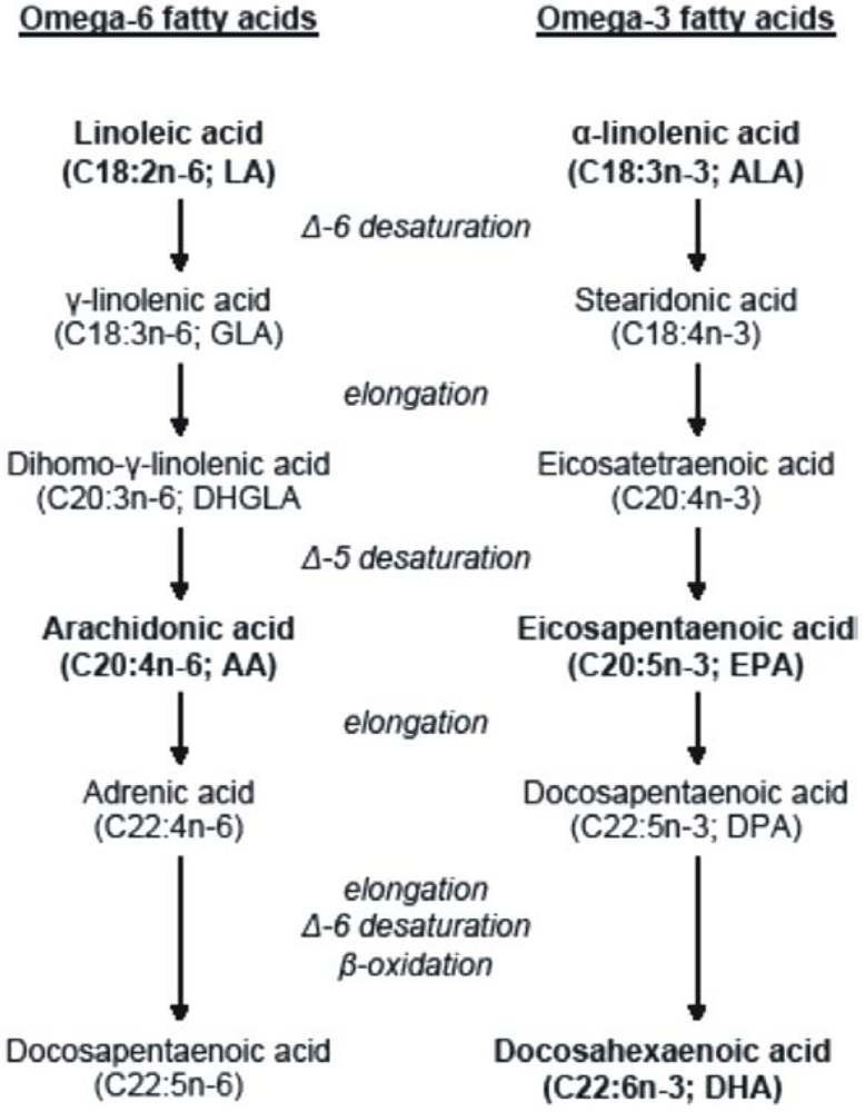
Mutta omega3- ja omega6 monityydytttämättömien rasvahappojen (PUFA) sarjan alkumolekyylejä tarvitaan valmiina elintarvikkeista. Niitä sanotaan sen takia essentielleiksi rasvahapoiksi (EFA). Nämä essentiellit rasvahapot linolihappo (C18:2 n6, LA) ja alfa-linoeelinihappo (C18:3 n3, ALA) muokataan sitten kehon oman aineenvaihdunnan entsyymeillä eteenpäin kumpikin omaa rataansa ( yksinkertaistetu kaava : https://res.mdpi.com/nutrients/nutrients-02-00965/article_deploy/html/images/nutrients-02-00965-g001-1024.png )
Niihin lisäytyy entsyymeillä ( desaturaasi) uusia kaksoissidoksia ja ne pidentyvät (elongaatio) usean kerran 2 hiilen pätkillä . Molemmat rasvahapot LA ja ALA käyttävät samoja desaturaasi- ja elongaasientsyymejä (= kilpailevat samoista entsyymeistä). Kun niiden johdannaiset ovat pitkäketjuista ( 24 hiiltä) niihin tulee vielä mutkaksi betaoksidaatiovaihe, jossa poistuu 2 hiiliketjua ja sitten vasta pysyy 6- kaksoissidoksen tärkeä 22:6 DHA n3- muoto paremmin pysymään.

( NNR2012 linkistä saa hyvän selityksen sivulta 220 , kuva 10.1) Siinä kuvassa on myös omega9 sarjan öljyhappo C18:1 näkyvissä linjoineen. Steariini happo C18:0 on SFA ja se voi saada kehossa jo yhden kaksoissidoksen ja muuttuu öljyhapoksi (18:1 n9) joten öljyhappo ei ole essentielli, mutta dietääristi suotuisa tähän tasapainokarttaan ja sillä on oma ratansa pidentyä.

Kasvien ja vapaana elävien kalojen tyydyttämättömät rasvahapot ovat pääasiassa cis-rasvahappoja.
  • The human body is capable of synthesising SFA and MUFA - including n-7 and n-9 series MUFA- from acetate, but n-3 and n-6 series PUFA are required from the diet. Linoleic acid ( n-6, LA) and alfa-linolenic acid (n-3, ALA) are metabolised ( desaturated and elongated) further in the body by the same enzyms. Naturally occurring unsaturated fatty acids in plants and wild fish are mainly cis-fatty acids.

(Karttakuva dieetin C18- kokoisten rasvahappojen aineenvaihdunnan radoista sivu 220)
2.6. 2018
SIVUMAININTA: Ajatuksia ÖLJYHAPOSTA
Päivitys: koetan etsiä tietoa öljyhapon (C18:1) metaboliakartasta . Se on mielestäni vähän epäselvä asia, enkä ole siihen kiinnittänyt enne huomiota. Kyselin mielessäni. Käyttääkö öljyhappo omega 3 tai omega6 linjan elongaasia tai desaturaasia ja arvelen, että ei ehkä käytä. Täytyy tarkistaa. Kovasti tulee isomeerejä siitä 18 - koosta.
Muistiin pari uutta artikkelia:https://www.ncbi.nlm.nih.gov/pubmed/29797206 (Kromi)
https://www.ncbi.nlm.nih.gov/pubmed/24823908 (metastasis) Gastric cancer and breast cancer have a clear tendency toward metastasis and invasion to the microenvironment predominantly composed

of adipocytes. Oleic acid is an abundant monounsaturated fatty acid that releases from adipocytes and impinges on different energy metabolism responses. The effect and underlying mechanisms of oleic acid on highly metastatic cancer cells are not completely understood. We reported that AMP-activated protein kinase (AMPK) was obviously activated in highly aggressive carcinoma cell lines treated by oleic acid, including gastric carcinoma HGC-27 and breast carcinoma MDA-MB-231 cell lines. AMPK enhanced the rates of fatty acid oxidation and ATP production and thus significantly promoted cancer growth and migration under serum deprivation. Inactivation of AMPK attenuated these activities of oleic acid. Oleic acid inhibited cancer cell growth and survival in low metastatic carcinoma cells, such as gastric carcinoma SGC7901 and breast carcinoma MCF-7 cell lines. Pharmacological activation of AMPK rescued the cell viability by maintained ATP levels by increasing fatty acid β-oxidation. These results indicate that highly metastatic carcinoma cells could consume oleic acid to maintain malignancy in an AMPK-dependent manner. Our findings demonstrate the important contribution of fatty acid oxidation to cancer cell function.

(2014)” The physiological roles of oleic acid in health and disease in humans are minimally investigated and understood. High-metastatic cancer cells and low metastatic cancer cells had opposite responses to oleic acid treatment with respect to cell survival and migration..”..”We concluded that high metastatic cancer cells with high-level consumption for energy utilise OA more efficiently than their counterpart cells”. (Öljyhappo edistää fosfatidyylikoliinin ja triglyseridin synteesiä*).
..
https://www.ncbi.nlm.nih.gov/pubmed/29730927J Agric Food Chem. 2018 May 23;66(20):5237-5246. doi: 10.1021/acs.jafc.8b01954. Epub 2018 May 10.
Epoxy Stearic Acid, an Oxidative Product Derived from Oleic Acid, Induces Cytotoxicity, Oxidative Stress, and Apoptosis in HepG2 Cells.
In the present study, effects of cis-9,10-epoxy stearic acid (ESA) generated by the thermal oxidation of oleic acid on HepG2 cells, including cytotoxicity, apoptosis, and oxidative stress, were investigated. Our results revealed that ESA decreased the cell viability and induced cell death. Cell cycle analysis with propidium iodide staining showed that ESA induced cell cycle arrest at the G0/G1 phase in HepG2 cells. Cell apoptosis analysis with annexin V and propidium iodide staining demonstrated that ESA induced HepG2 cell apoptotic events in a dose- and time-dependent manner; the apoptosis of cells after treated with 500 μM ESA for 12, 24, and 48 h was 32.16, 38.70, and 65.80%, respectively. Furthermore, ESA treatment to HepG2 cells resulted in an increase in reactive oxygen species and malondialdehyde (from 0.84 ± 0.02 to 8.90 ± 0.50 nmol/mg of protein) levels and a reduction in antioxidant enzyme activity, including superoxide dismutase (from 1.34 ± 0.27 to 0.10 ± 0.007 units/mg of protein), catalase (from 100.04 ± 5.05 to 20.09 ± 3.00 units/mg of protein), and glutathione peroxidase (from 120.44 ± 7.62 to 35.84 ± 5.99 milliunits/mg of protein). These findings provide critical information on the effects of ESA on HepG2 cells, particularly cytotoxicity and oxidative stress, which is important for the evaluation of the biosafety of the oxidative product of oleic acid.
(Hm- oljyhapolla ravinnon käristäminen ??)


onsdag 30 maj 2018

Tervetuloa sisään ja riisukaa takkinne!! ( Vaipallinen virus tuli sisään soluun ja riisui manttelinsa ja kävi taloksi.

Cell Microbiol. 2010 Oct;12(10):1378-88. doi: 10.1111/j.1462-5822.2010.01510.x. "Come in and take your coat off" - how host cells provide endocytosis for virus entry.
Abstract

VIRUKSET ovat solunsisäisiä loisia  ja  ne tarvitsevat isänsäsolun koneiston elinsykliisä ja virioniensa tuottoon.  Vasta kehkeytyneitten  viruspartikkeleiten  tehtävänä on välittää  genominen informaatio uusiin infektoitumattomiin soluihin ja organismeihin.  Viruksen sisäänmeno soluun pn se prosessi, jolla  se hankkii itselleen pääsyn   virusksen replikoitumiskohtiin infektoitumattoman solun sisällä, tämä on monivaiheinen tapahtumasarja, joka alkaa  kiinnittymisestä kohdesoluihin. Koska virukset ovat erittäin ykinkertaisia rakenteeltaan ja kokoomukseltaan ja niiltä puuttuu liikkumiskyky, ne tarvitsevat sadoittain isäntäsolun proteiineja  solun sisään pääsemiseen. (ENTRY)
  • Viruses are intracellular parasites that rely upon the host cell machinery for their life cycle. Newly generated virus particles have to transmit their genomic information to uninfected cells/organisms. Viral entry is the process to gain access to viral replication sites within uninfected cells, a multistep course of events that starts with binding to target cells.
  •  Since viruses are simple in structure and composition and lack any locomotive capacity, viruses depend on hundreds of host cell proteins during entry. Most animal viruses take advantage of endocytosis to enter cells.
 Solubiologiset , morfologiset ja biokemialliset tutkimukset, elävän solun kuvaukst ja systemaattiset lähestymistavat ovat auttaneet  tunnistamaan  erilaisia uusia endosyyttisiä mekanismeja  klatriinivälitteisen endosytoosin, makropinosytoosin ja kaveola/lipidilevy-välitteisen endosytoosin ohella. Sentakia  viruksen soluun menon tutkimus on muuttunut entistä monimutkaisemmaksi . tässä katsauksessa  on  biologinen yleisnäkymä  olemassa olevista  endosytoottisista mekanismeista ja strategioista, joita virukset käyttävät tai mahdollisesti käyttävät  solun sisäänmenossa.
  •  Cell biological, morphological and biochemical studies, live cell imaging and systematic approaches have identified various new endocytic mechanisms besides clathrin-mediated endocytosis, macropinocytosis and caveolar/lipid raft-mediated endocytosis. Hence, studying virus entry has become ever more complex. This review provides a cell biological overview of the existing endocytic mechanisms and strategies used or potentially used by viruses to enter cells.
  • PMID: 20678171
  • DOI: 10.1111/j.1462-5822.2010.01510.x
  •  Introduction

    Virus particles have a single purpose – to safely transfer their genomic information from infected to uninfected cells/organisms. Composed of nucleic acids (RNA or DNA), proteins and – for enveloped viruses membrane lipids, viruses are rather simple in structure and lack any metabolic or motile processes. Hence, they have evolved a ‘Trojan horse’ strategy to exploit the cell machinery for entry into target cells, a complex programme that involves hundreds of cellular proteins.
    During this process, two major tasks are fulfilled by the viruses through a series of complex interactions with the cell: (i) to overcome the obstacles that bar the virus from the site of replication such as the plasma membrane, a crowded cytoplasm and the nuclear membrane, and (ii) to release the genomic information at the right place within the cell to ascertain viral transcription and replication. Both tasks are coordinated in time and space and rely on a number of ‘cues’ provided by interactions of the host cell with the virus structure.
  • The virus particle

    During transmission, the viral genome is protected by a protein shell typically termed capsid. Capsids are mostly spherical, often icosahedral structures, which are composed of many subunits of one or more structural proteins. Enveloped viruses have an additional lipid membrane displaying viral membrane glycoproteins that engage receptors and mediate fusion events with cellular membranes (Harrison, 2008).
    Virus structures are stable – crosslinked by networks of intermolecular interactions to withstand hostile conditions encountered in the extracellular space. In fact, virus structures are metastable – poised upon cellular cues to undergo major conformational changes for an eventual release of the genomic information, a stepwise process termed uncoating. It is of fundamental importance that uncoating does not prematurely expose the viral genome, since this would lead to degradation and/or failed transport to the site of replication.

    Virus entry – an overview

    As viruses can only infect cells to which they can bind, cell receptors largely define the organism and cell tropism of a virus. Receptors promote entry by binding, by initiating conformational changes in the virus, by activating cellular signalling, and by inducing fusion at the plasma membrane or by promoting endocytosis. In fact, some viruses bind to several different receptors simultaneously or in sequence such as Human Immunodeficiency Virus type 1 (HIV‐1), adenoviruses and coxsackie B3 virus (Nemerow, 2000; Coyne and Bergelson, 2006;Mercer et al., 2010). Receptor molecules include a wide variety of different proteins, lipids and carbohydrates (for an overview see Helenius, 2007).
    After receptor binding, viruses undergo a period of diffusive or directed motion on the plasma membrane, until they become confined (Burckhardt and Greber, 2009). In addition, some viruses can move along filopodia to internalization sites by receptor interaction with the actin cytoskeleton and retrograde flow within filopodia (Lehmann et al., 2005; Schelhaas et al., 2008). 

    As mentioned, virus binding to receptors and ensuing signalling induce endocytosis and/or prepare the cell for invasion. While several enveloped viruses such as HIV or herpesviruses fuse directly with the plasma membrane to release the capsids into the cytoplasm, they also can use endocytosis depending on the cell system (Nicola et al., 2003; Miyauchi et al., 2009). In fact, most viruses use endocytosis, because this mode of entry is clearly advantageous: (i) endocytosis leaves no trace of the viral presence on the plasma membrane – likely to cause a delay of the immune response, (ii) endocytic uptake and ensuing vesicular transport provide a built‐in transport mechanism across the plasma membrane, the underlying actin cytoskeleton and the crowded cytoplasm, and (iii) vesicular trafficking provides access to intracellular organelles that allow viruses to ‘sense’ their environment by gradually changing conditions such as pH, redox environment and presence of specific proteases.
    For activation of membrane penetration or uncoating, structural changes in viruses are triggered by the host cell. While viruses can be already activated at the plasma membrane by, e.g. receptor binding as described above, activation also occurs often by the intraluminal environment of a particular organelle. The activated viruses then penetrate the vacuolar membrane delivering the viral structure or genome into the cytosol. Viral capsids often bind directly to microtubular motors for further cytosolic transport (Dohner and Sodeik, 2005). Most RNA viruses replicate in specific regions of the cytosol, whereas most DNA viruses must gain access to the nucleus. Nuclear import is typically achieved by cooperation with the nuclear import machinery (Whittaker et al., 2000).

    Endocytosis

    Endocytosis is essential in eukaryotic cells to internalize extracellular particles, fluid and ligands generally termed cargo (Conner and Schmid, 2003). Endocytosis starts with the formation of primary endocytic vesicles (PEVs), endocytic vacuoles generated by pinch‐offs from the plasma membrane. This multistep process involves activation, cargo capture/sorting, induction of membrane curvature, dilation of curvature and scission. PEVs are targeted to endosomal organelles. From there, cargo is further sorted to destination organelles.
    For a long time, it has been thought that receptor‐mediated endocytosis is mainly comprised of clathrin‐mediated endocytosis (CME). However, current research indicates a complex network of diverse continuing and triggered pathways (Fig. 1). In addition to CME, macropinocytosis and caveolar/raft‐dependent endocytosis, several further mechanisms have emerged that are less well characterized. They differ primarily in the formation of PEVs and involve a large number of cellular factors. The various endocytic phenomena will be briefly described (for more detail please refer to Gruenberg, 2001; Kirkham and Parton, 2005; Bonifacino and Rojas, 2006; Mayor and Pagano, 2007; Doherty and McMahon, 2009; Hansen and Nichols, 2009).
  • Endosomes

     Primary endocytic vesicles are routed to endosomes that are responsible for sorting, recycling, degradation, storage and transcytosis of cargo. The different endosomal organelles are heterogeneous in composition. They can fuse, some homo‐ others also heterotypically, and they undergo molecular changes with time. For simplicity, endosomal organelles are described here as collective identities, i.e. early endosomes (EE), late endosomes (LE), lysosomes and recycling endosomes (RE). EE are more dispersed throughout the cytosol, whereas RE, LE and lysosomes are located mainly perinuclearly, so that molecular sorting leads cargo such as viruses to different locations within the cell. Vesicular traffic and movement of endosomes occurs along microtubules. Cargo internalized by CME is typically delivered to EE 2–5 min after internalization, reach LE by 10–15 min and lysosomes by 30–60 min (Kielian et al., 1986; Mukherjee and Maxfield, 2004; Lakadamyali et al., 2006). Endosomes contain different membrane domains often specified by different Rab GTPases and their effectors, or phosphoinositides. Rab GTPases are prenylated proteins that associate with membranes and specifically localize to domains through their interactions with effector proteins (Pfeffer and Aivazian, 2004). Different Rab GTPases are associated with sorting of cargo to further destinations, e.g. Rab5 (EE), Rab4 (fast recycling to the plasma membrane), Rab11/22 (slow recycling by RE), Rab7 (LE), Rab9/retromer (trans‐GOLGI‐network, see Fig. 1). The presence of Rab GTPases and their effectors are also used for organelle specification (Zerial and McBride, 2001). Early endosomes are the first and main sorting station for cargo. From EE, cargo is transferred to RE, LE, or back to the plasma membrane. There are at least two variants of EE, highly motile and rather static ones that differ in the rate they convert to LE exemplifying the heterogeneity of endosome identity (Lakadamyali et al., 2006). Cargo sorting can be achieved by clustering of membrane proteins into domains. Membrane proteins that have been monoubiquitinated on the cytosolic side are sorted by the ESCRT complex into smaller vesicles that bud into the endosome lumen, forming intraluminal vesicles (ILV; Piper and Katzmann, 2007). This process starts in EE and leads to the multivesicular appearance of LE. The complex structure of EE, displaying vacuolar and long narrow tubular elements, likely directs diffusion of soluble and membrane proteins and could cause concentration gradients. Cargo destined for direct recycling is often concentrated in the tubular elements (Grant and Donaldson, 2009). The dimensions of ILV, and for viruses we can assume the same, mostly excludes entry into the tubular elements, so that they remain in the vacuolar part targeted for degradation. Most ILV are degraded in lysosomes. However, some backfuse with endosomal membranes. Vesicular stomatitis virus uses this feature to initially fuse with ILV, after which backfusion releases the capsid into the cytoplasm (Le Blanc et al., 2005). Endosome communication with the secretory pathway is provided by the retromer complex and Rab9 that allow vesicles to shuttle between endosomes and the trans‐GOLGI‐network (Bonifacino and Rojas, 2006). However, there is no clear evidence for viruses using the GOLGI route for entry. Instead, viruses mostly follow the route destined for degradation, from EE to LE to lysosomes. For degradation, cargo can be transported from EE to LE by vesicular transport. Alternatively, EE mature into LE (Rink et al., 2005; Vonderheit and Helenius, 2005). In the process, intermediate compartments also called maturing endosomes (ME) are established (Braulke and Bonifacino, 2009). ME exhibit markers for both, EE and LE. Endolysosomes form, when LE fuse with lysosomes. LE are ‘consumed’ in the process. Endolysosomes display a characteristic density in electron microscopy that increases when they condense into lysosomes (Mullock et al., 1998). From EE to lysosomes, the lumen of endosomes becomes increasingly acidic mainly through the action of V‐ATPase (Lafourcade et al., 2008). Also, the amount of hydrolases/proteinase increases by incoming transport from the GOLGI, so that viruses encounter a gradually changing chemical environment. A particular chemical environment provides the cues for structural changes in the particles by, e.g. a specific pH threshold and/or the activity of hydrolases/proteinases. Changes may trigger fusion of the viral envelope with endosomal membranes, partial disassembly of viruses, pore formation in endosomal membranes or more drastic changes such as rupture of endosomes by membrane destabilizing peptides. If a virus fails to penetrate the endosomal compartment of choice, its fate is usually degradation. It is noteworthy that endosomal maturation and vesicular transport are interconnected and interdependent, so that perturbation of a single function/factor may result in drastic consequences for several organelles, e.g. blocking V‐ATPase with bafilomycin A1 results not only in decreased intraluminal acidification of endosomes but also prevents formation of ME (Clague et al., 1994).


     Clathrin‐mediated endocytosis

     Clathrin‐mediated endocytosis is the endocytic pathway that is best understood (Doherty and Mcmahon, 2009). CME occurs constitutively in most mammalian cells. In addition, cargo such as viruses can also induce the formation of clathrin‐coated pits (CCPs) (Rust et al., 2004; Johannsdottir et al., 2009). For PEV formation, clathrin is recruited to the plasma membrane in response to internalization signals and forms a characteristic coat that is visible in electron microscopy. Many proteins are involved in coat assembly, although their functions remain in part elusive (Robinson, 2004). Typically, localized formation of phosphoinositide‐4,5‐phosphate (PI4,5P2) leads to recruitment of the adaptor protein AP2 that, in turn, recruits clathrin, AP180 and Eps15 followed by further adaptors involved in cargo selection/immobilization (Ungewickell and Hinrichsen, 2007). However, it has become clear that CCPs have not always the same composition (Robinson, 2004). AP2, for example, is, contrary to the early model, not always required for CME (Motley et al., 2003). The varying requirement for adaptor proteins indicates a common mechanism combined with a diverse degree of regulation. Fission of clathrin‐coated vesicles from the plasma membrane involves the GTPase dynamin (Hinshaw, 2000). From recruitment of clathrin to scission, PEV formation is fast and takes about 1 min for individual pits. Then, synaptojanin, auxilin and HSC70 help to disassemble the clathrin coat from the vesicle. PEVs are transported to EE. The first virus described to enter cells via CME was Semliki Forest Virus (SFV) (Helenius et al., 1980). Many other viruses from diverse virus families use CME for entry including adenovirus 2 and 5, hepatitis C virus, dengue virus and influenza A virus (Mercer et al., 2010). In addition to entry by CME, viruses may also use alternate pathways in the same cell, at the same time, as has been described for influenza A viruses (Rust et al., 2004).

    Caveolar/raft‐mediated endocytosis 

     The term caveolar/raft‐mediated endocytosis will be applied to caveolar and caveolin‐independent mechanisms of endocytosis that are lipid raft‐dependent (Pelkmans et al., 2001; Pelkmans et al., 2002; Parton and Richards, 2003; Damm et al., 2005). As suggested by Kirkham and Parton (2005), they may share a congeneric core mechanism somewhat similar to CME requiring different sets of molecular factors. Characteristically, formation of PEVs requires lipid rafts and tyrosinekinase/phosphatase‐regulated, ligand‐triggered signalling events. Uptake can involve caveolae and dynamin‐2, but may occur without. While it has been previously thought that a novel organelle, the caveosome, acts as an intermediate station in intracellular trafficking, recent work from the Helenius laboratory indicates that this organelle corresponds to modified late endosomes/lysosomes (Mercer et al., 2010; A. Helenius, pers. comm.). From there, cargo is often routed to the endoplasmic reticulum (ER). How transit from endosomes to the ER occurs is not entirely clear yet, but the process seems to involve lipid‐sorting events (Qian et al., 2009). Cellular cargo includes glycolipids and some glycosyl‐phosphatidylinositol‐(GPI)‐anchored proteins and their ligands (Lajoie and Nabi, 2007). Studying the entry of polyomaviruses has contributed much to our understanding of this pathway. Polyomaviruses such as Simian Virus 40 (SV40) and mouse polyomavirus (mPy) are small non‐enveloped DNA viruses with capsids assembled from 72 pentamers of the major capsid protein VP1. Polyomaviruses use different gangliosides as receptors. As an example, SV40 associates with detergent‐resistant microdomains in the plasma membrane and enter uncoated, tight‐fitting pits (Kartenbeck et al., 1989; Damm et al., 2005). Caveolae and pits formed in caveolin‐deficient cells are morphologically indistinguashable. Internalization and vesicular trafficking is asynchronous and slow, e.g. SV40 reaches the ER roughly 6 h post infection (Kartenbeck et al., 1989). Internalization is inhibited by cholesterol depletion, by inhibitors of actin dynamics and by inhibitors of tyrosine kinases (Pelkmans et al., 2002; Damm et al., 2005). Conversely, internalization is accelerated by inhibitors of serine/threonine or tyrosine phosphatases. RhoA seems to regulate the actin polymerization events. SV40 entry into caveolin‐deficient cell lines is mechanistically similar to entry by caveolar endocytosis but does not require dynamin‐2 (and obviously not caveolin‐1), and is less dependent on actin dynamics (Damm et al., 2005). Internalization is also faster in caveolin‐deficient cells suggesting that caveolin/dynamin introduce an additional level of regulation. Depending on the cellular system, it is likely that both variants of caveolar/lipid raft‐mediated endocytosis operate in parallel.
      

                Macropinocytosis/phagocytosis      

    Macropinocytosis and phagocytosis differ from other endocytic mechanisms in that they require extensive actin cytoskeletal reorganization. These rearrangements are coupled to an outward‐directed formation of plasma membrane extensions, whereas in other pathways the plasma membrane ‘buds’ into the cell. Under normal conditions, macropinocytosis is induced by growth factors. Depending on cell type and stimulation, formation of membrane ruffles, filopodia, or ‘bleb’ formation precedes the generation of macropinosomes. Macropinosomes form by backfolding of membrane extensions and fusion with the plasma membrane (Swanson and Watts, 1995). Depending on the extent of membrane extension, resulting macropinosomes vary in shape and size. With diameters of up to 10 µm, they are relatively large. Macropinocytosis transiently increases fluid uptake up to 10‐fold. The activation of macropinocytosis and formation of PEVs requires sodium/proton exchanger, the RhoGTPases Rac1 and/or Cdc42, various cellular kinases (e.g. PAK1, PKC), CtB1 and cholesterol (Kerr and Teasdale, 2009; Mercer and Helenius, 2009). There is a significant degree of cell type dependence for further requirements such as the GTPases Rab34, Arf6 or actin modulatory factors. Macropinosomes can be recycled to the plasma membrane, undergo intraluminal acidification, homotypic fusion and heterotypic fusion with EE (Racoosin and Swanson, 1993; Hewlett et al., 1994; Swanson and Watts, 1995). Viruses that use macropinocytosis for entry include, e.g. vaccinia virus and Kaposi's sarcoma‐associated herpesvirus (Mercer and Helenius, 2009; Raghu et al., 2009). Another group of viruses induces macropinocytosis and requires it for entry, yet does not use it for internalization. This group includes, e.g. species C human adenoviruses 2 and 5 and rubella virus. For adenovirus 2, macropinocytosis is required for penetration of endosomal membranes after CME (Meier et al., 2002). The mechanism is not entirely understood but seems to require lysis of macropinosomes.


     Phagocytosis is not commonly used for viral entry but for uptake of large particles such as bacteria. Actin rearrangement and protuberance of the plasma membrane is induced and guided by an external particle bound to the cell. A tight fitting endocytic vacuole is formed around the particle with no or little fluid uptake (Swanson, 2008). Factors commonly involved include actin, RhoA, tyrosine kinases, cholesterol and dynamin‐2. A number of cell type‐dependent factors can be required, e.g. AP2, Arf6, Cdc42 and Rac1. The entry of mimivirus, an amoebal pathogen, and the entry of herpes simplex virus into corneal fibroblasts were found to be consistent with phagocytosis (Clement et al., 2006; Ghigo et al., 2008). For the purpose of this review, further novel endocytic pathways can be subdivided into (i) pathways described cell biologically but not for virus entry, and (ii) virus entry pathways by endocytic mechanisms inconsistent with any of the above. Both have in common that we have to learn much more about them to understand their use and their relationship to other pathways, e.g. whether some may share similar core mechanisms with a variation of factors. Novel cell biologically described pathways The internalization of GPI‐anchored proteins such as GPI‐GFP, decay‐accelerating factor and folate receptor GPI occurs by tubular endocytic pits resulting in the generation of ‘GPI‐anchored enriched endocytic compartments’ (GEECs; Sabharanjak et al., 2002). GEEC endocytosis requires lipid rafts but not clathrin, caveolin or dynamin. During pit formation, the GTPase Arf1 is recruited to the plasma membrane (Kumari and Mayor, 2008). Arf1 recruits ARHGAP10, a GAP for Cdc42, which regulates and precedes internalization of cargo by actin polymerization dynamics. GRAF1 may be a coat factor. The resulting GEEC, a tubular PEV, is not readily observed, since it is fragile and very sensitive to fixation conditions (Kirkham et al., 2005). Subsequent fusion of GEECs with the early endosome occurs in a Rab5/PI3 kinase‐dependent manner (Kalia et al., 2006). Flotillin‐1 has been implicated in the internalization of GPI‐anchored proteins (e.g. CD59), cholera toxin B and proteoglycans (Glebov et al., 2006; Payne et al., 2007; Hansen and Nichols, 2009). Flotillins are membrane‐bound, ubiquitously expressed, highly conserved proteins that share a similar membrane topology with caveolins. Flotillin‐1 heterooligomerization with flotillin‐2 to form microdomains at the plasma membrane depends on cholesterol and is required for assembly and function (Babuke et al., 2009). Internalization by flotillin microdomains is regulated by fyn kinase. The resulting flotillin‐1‐positive PEV fuses with EE. It is noteworthy that flotillin‐1 depletion may result in decreased or increased caveolin‐1 levels depending on the cell type by a non‐transcriptional mechanism (Vassilieva et al., 2009, M. Schelhaas and A. Helenius, unpubl. results). Endocytosis of MHCI and CD59 into HeLa cells is sensitive to cholesterol depletion and is associated with membrane ruffling initiated by the GTPase Arf6, the defining factor for this pathway (Naslavsky et al., 2004). Arf6 recruits and stimulates PI5 kinase to produce PI4,5P2 (Brown et al., 2001). After internalization, PI4,5P2 hydrolysis occurs and results in an Arf6‐positive compartment that fuses with the early endosome 5–10 min after PEV formation (Naslavsky et al., 2003). It is noteworthy that this pathway may be specific to HeLa cells, as Arf6 has been additionally implicated in macropinocytosis and CME for other cargos and/or cell types. The uptake of IL‐2 and gamma‐c cytokine receptor is independent of clathrin but occurs by a dynamin‐dependent process (Lamaze et al., 2001; Sauvonnet et al., 2005). IL‐2 binding to its receptor leads to prominent association of the receptor with lipid rafts, which is coupled to internalization by non‐coated, non‐tubular pits. Actin polymerization dynamics regulated by RhoA are also required for internalization. Cortactin – a potential cofactor for CME – participates in this process independently of clathrin. The PEV is routed to EE. Viruses that use unusual endocytic pathways Lymphocytic choriomeningitis virus (LCMV) entry illustrates an endocytic pathway that has not been observed previously, but which has been defined on negative terms. LCMV, an arenavirus, uses alpha‐dystroglycan as major receptor for cell entry (Cao et al., 1998). Endocytosis of LCMV occurs in smooth, non‐coated pits (Quirin et al., 2008). Internalization is largely independent of clathrin, caveolin, dynamin, actin dynamics and lipid rafts but requires membrane cholesterol (Quirin et al., 2008; Rojek et al., 2008). LCMV internalization is also independent of Arf6 and flotillin‐1. After internalization, LCMV particles mostly bypass EE and are routed to LE directly from the plasma membrane (Quirin et al., 2008). Entry of Human papillomavirus type 16 (HPV‐16), a non‐enveloped virus of the papillomavirus family, occurs likewise by a novel endocytic mechanism. Virus particles consist of two structural proteins (L1, L2) that form an icosahedral (T = 7) particle of about 55 nm in diameter. After an initial interaction with heparan sulfate proteoglycans followed by a sequence of structural changes, the virus is released and transferred to an unknown co‐receptor (Selinka et al., 2007; Sapp and Day, 2009). Endocytosis occurs by a clathrin‐, caveolin‐, flotillin‐, lipid raft‐, dynamin‐independent mechanism that is distinct from macropinocytosis and phagocytosis (Spoden et al., 2008; M. Schelhaas et al., unpublished). Actin polymerization events independent of classical Rho‐like GTPases drive scission of wide, uncoated pits of up to 100 nm in diameter leading to PEV formation. Endocytosis further depends on PI3 kinase, protein kinase C and sodium/proton exchange (M. Schelhaas et al., unpublished). Intracellular trafficking occurs through endosomes. Why did viruses evolve to use a particular pathway? With multiple endocytic pathways to choose from, viruses must have evolved to follow a particular route for specific reasons. What are these? To date, very little evidence exists to substantiate answers to this question. One simple reason may be the standard size of endocytic pits/PEVs. Clathrin‐coated pits are somewhat variable in size and can accommodate larger cargo, and the same may be true for other pathways. However, large viruses such as herpesviruses, poxviruses and mimivirus with particles bigger than 150 nm in diameter may have evolved to use macropinocytosis or phagocytosis that easily provide a big enough container for such a large cargo. Another trivial but unsubstantiated reason may be that the primary target cells/tissues for certain viruses more actively deploy a certain endocytic mechanism. However, some evidence for a role of different endocytic organelles in virus entry is provided by the viruses' necessity for membrane penetration and uncoating. Many enveloped viruses require a low intra‐endosomal pH for activation of the fusiogenic activity of the viral glycoproteins to release their capsids into the cytoplasm. Depending on the pH threshold and the intraluminal pH of endosomes, they likely fuse in EE (e.g. VSV, SFV; Marsh et al., 1983; Johannsdottir et al., 2009), ME/LE (e.g. influenza A; Matlin et al., 1981; 1982), or potentially endolysosomes/lysosomes (e.g. vaccinia; Mercer et al., 2008). LCMV may have chosen a route that mostly bypasses EE for a quick rather than a gradual drop in pH, when it is delivered to LE (Quirin et al., 2008). Similarly, non‐enveloped viruses can be pH‐dependent for membrane penetration and uncoating. In addition, viruses may require a low pH in endosomes for the activity of pH‐dependent proteases within endosomes that results in shedding of membrane penetration factors/partial uncoating. In particular cathepsins, endosomal cysteine proteases, were found to proteolytically cleave viral surface proteins of a variety of different viruses/virus families (e.g. Golden et al., 2004; Chandran et al., 2005; Simmons et al., 2005; Schornberg et al., 2006; Diederich et al., 2008; Regan et al., 2008). Most of the proteolytically or pH‐induced structural changes in endosomes occur within minutes, once the threshold has been reached. In contrary, HPV‐16 requires hours in the endosomes for membrane penetration (M. Schelhaas et al., unpubl. results). Hence, we have to learn much more about the kinetics of endocytic events, about timing or retaining of virus trafficking in combination with the associated structural consequences for viruses to understand the role of particular organelle environments in entry. Polyomaviruses such as SV40 and mPy seem to use caveolar/raft‐dependent endocytosis, since it can lead cargo to the endoplasmic reticulum. There, the viruses use molecular chaperones of the biosynthetic machinery to partially uncoat: ERp57 isomerizes intermolecular disulfide bonds in the SV40 capsid to release specifically the vertex capsomers (Schelhaas et al., 2007), ERp29 externalizes the stabilizing C‐terminal arm of the major capsid protein VP1 from intercapsomer connections in mPy (Magnuson et al., 2005). In addition, both seem to use the ER‐associated degradation machinery for transport across the ER membrane to the cytosol for eventual import into the nucleus (Lilley et al., 2006; Schelhaas et al., 2007). The viruses most likely have evolved to use the endocytic route to the ER, since the required factors/functions are unique to this organelle. Finally, a largely speculative thought deals with viral requirements for signal transduction events. As mentioned, virus endocytosis often requires signal transduction. In a systematic siRNA screen for the kinase requirements of VSV and SV40 endocytosis, a high number (208 of 590) was found to regulate the entry of these viruses (Pelkmans et al., 2005). Many of the kinases directly fine tune endocytic events or are important for a certain cell state that allows infection (e.g. migratory versus non‐migratory cells; Snijder et al., 2009). In addition, it is likely that several of the required kinases are needed for viral signal transduction events to prepare the cell for invasion and/or omit anti‐viral responses such as apoptosis. In general, signalling during endocytosis is compartmentalized (Sorkin and von Zastrow, 2009). It can occur at the plasma membrane, it may occur at the plasma membrane and from within endosomes (sustained signalling; Vieira et al., 1996), or it may occur solely from within endosomes (Daaka et al., 1998) depending on the cell system or signalling components. This compartmentalization of signalling provides for a variety of cellular effects that viruses may take advantage of by choosing a particular endocytic pathway. Conclusions Studying virus endocytosis is as complex as the choice of endocytic mechanisms/vesicular traffic and their numerous factors for entry are. An additional level of complexity is provided by existing and potential interconnections and interdependences between endocytic phenomena. The complexity increases even further, since viruses potentially use multiple routes in different cell types and/or within a single cell. Also, it is now clear that low pH‐activated viruses do not necessarily use CME, so that for some viruses it may be required to carefully re‐evaluate their entry pathway. Several considerations need to be made when studying the cell biology of virus endocytosis: virus inocula may contain only a small fraction of particles that enter productively. Microscopic or biochemical methods to follow viruses on their way into the cell do not allow a distinction between productive and non‐productive particles, so that investigations have to be complemented by techniques relying on infection. Cellular perturbation methods include the use of inhibitors, dominant negative mutants, small interfering RNA‐mediated silencing and mutant cells lines. As some of these cellular perturbations cause drastic changes in endocytosis beyond the intended, analysis of the perturbation effects must include morphological/biochemical techniques, and a careful use of cell biological and/or viral controls. Again, only a combination of biochemical, morphological, visual and functional methods allow the pathway(s) of entry to be analysed with confidence. Since viruses potentially use several different endocytic entry mechanisms in different cells or even a single cell, it will be necessary to relate the information obtained in tissue culture systems to in vivo infection models. This will be one of the major challenges for the future, as these model systems may be not available for certain viruses, and as cell biological perturbation studies in these model systems can be extremely difficult. Studies on virus endocytosis can provide new insights into endocytic mechanisms, as viruses can be easily followed by morphological techniques and provide access to reliable endocytosis endpoint detection methods, i.e. infection. These features are already exploited by siRNA screens aimed to determine host cell factors of virusinfections (e.g. Brass et al., 2008; Konig et al., 2008; Zhou et al., 2008; Li et al., 2009; Karlas et al., 2010). The information obtained may in addition to valuable information on the cell biology of virus infections provide new targets for antiviral therapy that would target the host instead of the virus. Acknowledgements

    måndag 30 april 2018

    Samassa geenissä kuin TRIM47 on myös ACOX

    Olen katsomasas kromosomin 17 TRIM-proteiineja.
    kromosomilocus 17q25.1,  missä sijaitsee  TRIM47 onkin  sellainen että  siihen sijoittuu tietty joukko   kiinnostusta herättävää geeniä.
    Yksi näistä koskee rasvahappoaineenvaihduntaa ja on peroksisomaalinen:  ACOX1, PALMCOX on  betaoksidaatiossa  tärkeä.
    http://www.genecards.org/cgi-bin/carddisp.pl?gene=ACOX1
    Otan talteen sen nimet ja vähän tietoa siitä:

    Entrez Gene Summary for ACOX1 Gene

    • The protein encoded by this gene is the first enzyme of the fatty acid beta-oxidation pathway, which catalyzes the desaturation of acyl-CoAs to 2-trans-enoyl-CoAs. It donates electrons directly to molecular oxygen, thereby producing hydrogen peroxide. Defects in this gene result in pseudoneonatal adrenoleukodystrophy, a disease that is characterized by accumulation of very long chain fatty acids. Alternatively spliced transcript variants encoding different isoforms have been identified. [provided by RefSeq, Jul 2008]

    GeneCards Summary for ACOX1 Gene

    ACOX1 (Acyl-CoA Oxidase 1) is a Protein Coding gene. Diseases associated with ACOX1 include Peroxisomal Acyl-Coa Oxidase Deficiency and Adrenoleukodystrophy. Among its related pathways are Estrogen Receptor Pathway and Peroxisomal lipid metabolism. Gene Ontology (GO) annotations related to this gene include receptor binding and protein N-terminus binding. An important paralog of this gene is ACOX2.

    UniProtKB/Swiss-Prot for ACOX1 Gene

    • Catalyzes the desaturation of acyl-CoAs to 2-trans-enoyl-CoAs. Isoform 1 shows highest activity against medium-chain fatty acyl-CoAs and activity decreases with increasing chain length. Isoform 2 is active against a much broader range of substrates and shows activity towards very long-chain acyl-CoAs. Isoform 2 is twice as active as isoform 1 against 16-hydroxy-palmitoyl-CoA and is 25% more active against 1,16-hexadecanodioyl-CoA.

    Aliases for ACOX1 Gene

    • SCOX 3 4
    • AOX 3 4
    • Peroxisomal Acyl-Coenzyme A Oxidase 1 3
    • Peroxisomal Fatty Acyl-CoA Oxidase 3
    • Acyl-CoA Oxidase, Straight-Chain 3
    • PALMCOX 3
    • Acyl-CoA Oxidase 1 2 3 5
    • Palmitoyl-CoA Oxidase 2 3 4
    • Acyl-Coenzyme A Oxidase 1, Palmitoyl 2 3
    • Straight-Chain Acyl-CoA Oxidase 3 4
    • Acyl-CoA Oxidase 1, Palmitoyl 2 3
    • EC 1.3.3.6 4 56
    • ACOX 3 4
    • TARKISTAN missä geenissä sijaitsee ACOX2 ja mitä se tekee.  Ei ainakaan samassa geenissä, vaan kromosomissa 3p14.3.

    •  http://www.genecards.org/cgi-bin/carddisp.pl?gene=ACOX2
    • Aliases for ACOX2 Gene

      • Acyl-CoA Oxidase 2 2 3 5
      • 3-Alpha,7-Alpha,12-Alpha-Trihydroxy-5-Beta-Cholestanoyl-CoA 24-Hydroxylase 2 3 4
      • Trihydroxycoprostanoyl-CoA Oxidase 2 3 4
      • 3-Alpha,7-Alpha,12-Alpha-Trihydroxy-5-Beta-Cholestanoyl-CoA Oxidase 3 4
      • Acyl-Coenzyme A Oxidase 2, Branched Chain 2 3
      • Acyl-CoA Oxidase 2, Branched Chain 2 3
      • Branched Chain Acyl-CoA Oxidase 2 3
      • THCA-CoA Oxidase 3 4
      • THCCox 3 4
      • Peroxisomal Branched Chain Acyl-CoA Oxidase 3
      • Peroxisomal Acyl-Coenzyme A Oxidase 2 3
      • EC 1.17.99.3 4
      • BRCACOX 3
      • BRCOX 3
      • CBAS6 3
      • BCOX 3